
FAU VIP Seed Grant
Quantum-Accelerated Discovery of Small Molecule Inhibitors Targeting Precursor Mitochondrial microRNAs in Cancer
PI: Ilyas Yildirim · Co-PIs: Shailaja Allani and Ajay Bommareddy · 2026–2027
$46,500
Disease-associated repeat RNAs adopt dynamic structures that can be recognized by carefully designed small molecules.
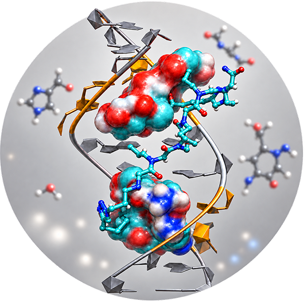
RNA folds into complex three-dimensional structures that contain internal loops, bulges, and other noncanonical motifs. These structural features can create selective binding pockets for small molecules. In disease-associated repeat RNAs, such motifs may contribute to abnormal RNA–protein interactions, toxic RNA accumulation, and disrupted cellular function. Small molecules that recognize these structures can potentially alter RNA behavior, block harmful interactions, or promote degradation of disease-causing RNA. This makes structured RNA an increasingly important target for the development of disease-modifying therapeutics.
This molecular model illustrates a small-molecule ligand targeting an expanded CCUG-repeat RNA, the pathogenic repeat RNA associated with Myotonic Dystrophy Type 2 (DM2). The visualization highlights how a ligand can recognize and interact with structural motifs formed by the CCUG repeats.
Accurate RNA simulations depend on force fields that reproduce stacking, torsions, and noncanonical interactions with high fidelity.
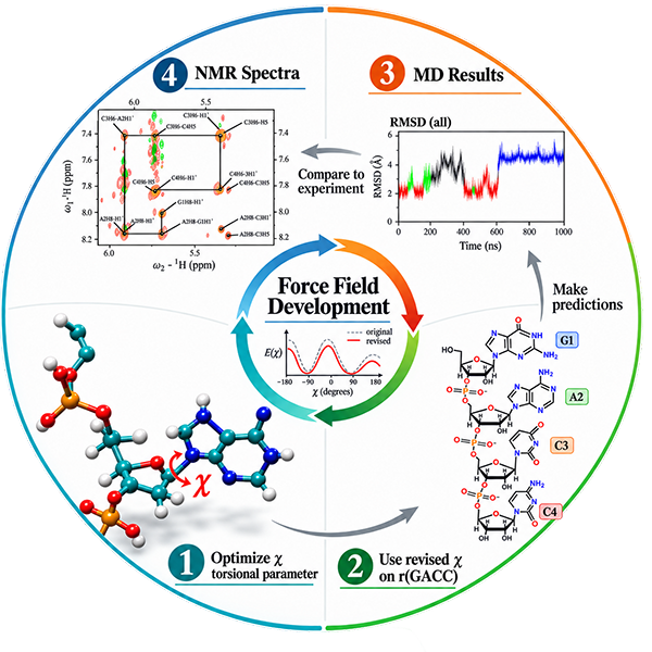
Molecular dynamics simulations describe biomolecular motion using a force field, a mathematical model that determines how atoms interact and how the molecular energy changes as bonds, angles, torsions, electrostatic interactions, and nonbonded contacts evolve. For RNA, the quality of these parameters is critical: even small inaccuracies can distort base stacking, backbone conformations, hydrogen bonding, and the relative stability of alternative structures. A carefully validated force field allows simulations to generate physically meaningful atomistic trajectories, revealing structural fluctuations and conformational transitions at a level of spatial and temporal detail that is difficult to obtain directly from experiments alone.
This figure highlights torsional degrees of freedom in nucleic acids, which strongly influence RNA backbone geometry and the orientation of nucleobases. Torsional parameters determine the energetic preference for different rotational states and therefore directly affect the structures sampled during molecular dynamics simulations. Research by Dr. Yildirim and collaborators included re-parameterization of several RNA torsions and extensive benchmarking against experimental NMR and thermodynamic data, leading to improved AMBER descriptions of RNA structure and energetics.
RNA sequence can encode many competing three-dimensional conformations. The lab approaches folding as an energy-landscape problem.
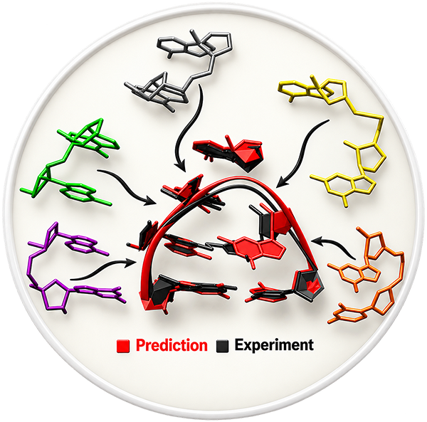
Predicting the three-dimensional structure of RNA directly from sequence remains one of the central challenges in computational structural biology. Unlike simple polymers, RNA molecules can adopt highly diverse folds stabilized by base pairing, base stacking, noncanonical interactions, backbone flexibility, solvent effects, and ion-mediated stabilization. Because RNA structure is tightly linked to function, the ability to predict RNA 3D architecture de novo would significantly expand our ability to understand gene regulation, RNA-mediated disease mechanisms, and the structural basis of therapeutic targeting. Reliable RNA structure prediction could therefore help identify new biological hypotheses and support the design of molecules that recognize disease-relevant RNA motifs.
This figure represents an RNA structure-prediction workflow centered on TAPERSS, a fragment-assembly framework for predicting RNA three-dimensional structures from sequence. In this approach, structural fragments and physical principles are combined to generate candidate folds, evaluate their plausibility, and refine structural models. The broader goal is to improve de novo RNA structure prediction and help bridge the gap between RNA sequence, structure, dynamics, and biological function.
Internal loops are dynamic structural elements that shape RNA folding, flexibility, and molecular recognition.
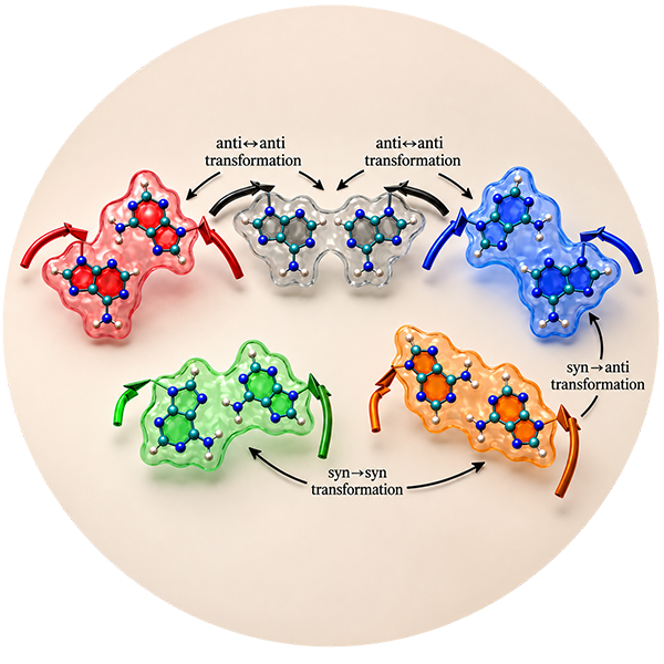
RNA internal loops are not static structural defects; they are dynamic motifs that can sample multiple conformations and reorganize in response to sequence context, ions, ligands, and protein binding. These motions are often central to RNA function because internal loops can shape local geometry, regulate tertiary contacts, influence folding pathways, and create recognition sites for proteins or small molecules. In some disease-associated RNAs, internal loops formed by repeat expansions contribute directly to pathology by promoting abnormal RNA structure and misregulating molecular recognition. Understanding the conformational landscapes of RNA internal loops is therefore important not only for fundamental RNA biophysics, but also for developing strategies to selectively target disease-relevant RNA motifs.
This image shows 1×1 AA RNA internal loops and highlights their dynamic behavior as characterized using advanced sampling techniques. Such methods are especially valuable because they can reveal low-population conformations and transitions between structural states that may be difficult to capture with conventional simulations alone. Mapping these conformational ensembles helps define how internal loops behave, how they contribute to RNA structure and function, and how they may be recognized selectively in RNA-targeted therapeutic design.
The lab also investigates broader nucleic-acid systems, from DNA deformation and transcription-factor complexes to programmable DNA/RNA molecular assemblies.
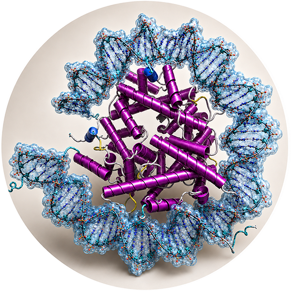
The nucleosome core particle is the fundamental repeating unit of chromatin in eukaryotic cells. It consists of DNA wrapped around a histone octamer composed of H2A, H2B, H3, and H4 proteins. This organization is essential for packaging the genome, but it also plays a central role in regulating DNA accessibility for transcription, replication, recombination, and repair. Because nucleosomal DNA is sharply bent and constrained around the histone core, its structure differs substantially from free DNA. Understanding how DNA accommodates this strong bending is important for explaining chromatin organization, gene regulation, and the molecular basis of disease-associated chromatin dysfunction.
This image shows a nucleosome core particle, in which DNA is wrapped around the histone octamer in a superhelical path. The visualization highlights the pronounced bending and packaging of DNA required for nucleosome formation. Such structural distortions are biologically important because they influence chromatin stability, nucleosome positioning, and the ability of regulatory proteins to access DNA. Abnormal nucleosome architecture or disease-associated histone mutations can alter these properties and thereby affect gene expression and genome maintenance.
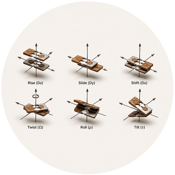
The geometry of DNA can be described using a compact set of structural parameters derived from the arrangement of neighboring Watson–Crick base pairs. In canonical DNA, adenine pairs with thymine and guanine pairs with cytosine, forming stacked base pairs along the double helix. The relative position and orientation of adjacent base pairs can then be quantified using base-pair and base-pair-step parameters such as shift, slide, rise, tilt, roll, and twist. These descriptors provide a simple but powerful framework for translating complex three-dimensional DNA structures into measurable geometric quantities that can be compared, modeled, and analyzed computationally.
This image illustrates the geometric parameters used to describe individual base pairs and the relationships between neighboring base-pair steps in DNA. By reducing complex DNA structures to a set of translational and rotational variables, researchers can quantify local distortions and identify patterns of bending along the double helix. Such descriptions are particularly useful for developing models and algorithms that analyze the strong DNA deformation required for wrapping around histone proteins in nucleosome core particles.
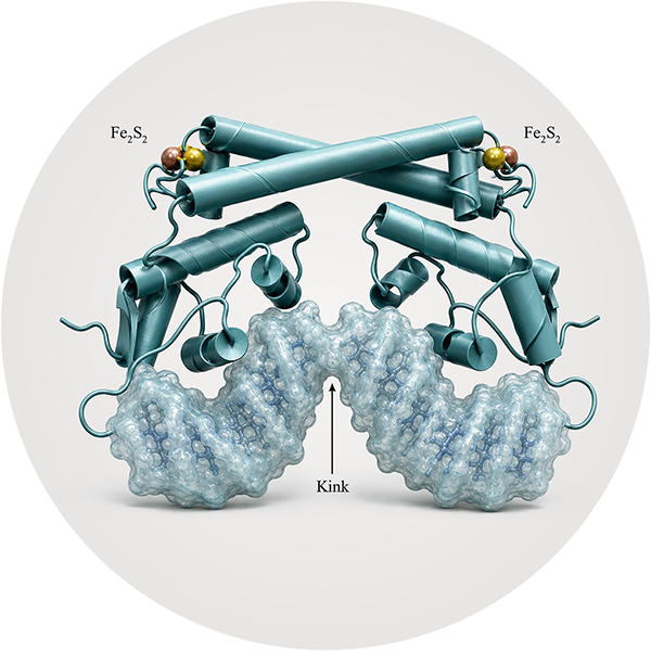
Transcription factors regulate gene expression not only through direct molecular recognition of DNA, but also by altering DNA structure itself. Protein binding can bend, kink, twist, or locally deform the double helix, changing the spatial arrangement of promoter elements and their accessibility to the transcriptional machinery. These structural changes can determine whether RNA polymerase recognizes a promoter efficiently and therefore whether a gene is activated or repressed. Understanding protein-induced DNA deformation provides an important structural framework for explaining how regulatory signals are transmitted through DNA geometry to control gene expression.
This image illustrates a transcription-factor–DNA complex and the structural deformation that can accompany protein binding. Related work by Yildirim and collaborators demonstrated that the metal-sensing transcription factor CueR can regulate transcription by changing the topology of its core promoter. In the repressor state, CueR slightly bends the promoter DNA in a way that prevents optimal recognition by RNA polymerase. Upon metal binding, CueR switches to an activator conformation and introduces torsional stress that kinks and undertwists the promoter DNA, reorganizing its geometry into a more favorable substrate for transcription. This work demonstrates how changes in DNA topology can transmit an allosteric regulatory signal and control gene expression.
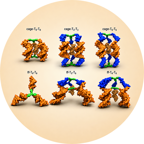
Small molecule–DNA hybrids combine the programmable molecular recognition of DNA with the chemical and structural properties of synthetic organic molecules. By covalently connecting hydrophobic organic cores to DNA strands, these hybrid building blocks can self-assemble in aqueous environments into well-defined nanoscale architectures. The structures that form depend not only on DNA hybridization, but also on the geometry of the organic core, linker flexibility, attachment direction, and the ability of hydrophobic surfaces to become shielded from water. Understanding how these molecular design parameters control self-assembly provides a framework for engineering programmable DNA-based nanostructures with predictable geometries and properties.
This image illustrates the self-assembly of small molecule–DNA hybrid building blocks into nanoscale structures. Work by Yildirim and collaborators demonstrated that even the direction in which an organic core is attached to DNA can strongly influence the resulting architecture. When the cores are connected to the 3′ ends of the DNA strands, favorable interactions can allow the cores to associate with the minor groove of the assembled duplex and promote cyclic dimer formation. Changing the linkage to the 5′ direction can alter these interactions and favor higher-order cyclic structures. These results demonstrate how subtle molecular design choices can control DNA-directed self-assembly.
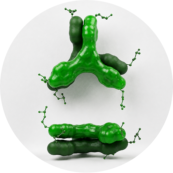
Hydrophobic interactions can provide a powerful driving force for the organization and stabilization of small molecule–DNA assemblies in aqueous environments. When hydrophobic organic cores are attached to DNA, the system tends to minimize their exposure to water through favorable interactions with neighboring organic cores, DNA bases, or base pairs. In appropriately designed assemblies, direct face-to-face association between the organic cores can strongly stabilize the resulting structure. These interactions work together with DNA hybridization to control molecular organization, thermal stability, and cooperative assembly, illustrating how noncovalent interactions can be deliberately exploited in the design of nucleic-acid-based nanomaterials.
This image illustrates the close association of hydrophobic organic cores within a small molecule–DNA hybrid assembly. Computational and experimental studies by Yildirim and collaborators showed that when the exposed hydrophobic surfaces of these cores are effectively minimized, favorable face-to-face core interactions can form and substantially stabilize the assembled structure. The organic cores can also interact with nearby DNA bases and base pairs, creating a network of hydrophobic and molecular contacts. Such well-organized assemblies display enhanced thermal stability and cooperative melting, demonstrating how molecular-scale core interactions can control the properties of DNA-based nanostructures.
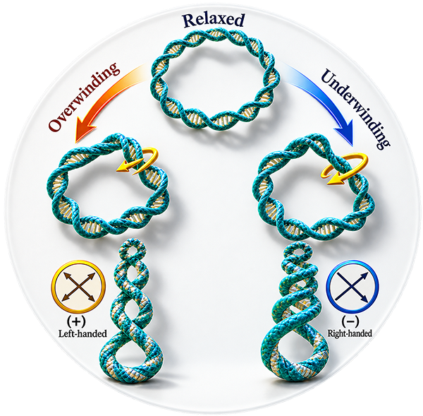
DNA is not always organized as a relaxed linear double helix. In cells, it is frequently bent, twisted, and supercoiled into higher-order structures that help compact the genome and regulate its accessibility. Superhelical organization arises when torsional strain changes the winding of the DNA double helix, causing the DNA axis itself to coil in space. These structural changes can influence transcription, replication, recombination, and DNA repair by altering local geometry and the energetic cost of opening or deforming DNA. Understanding how local base-pair geometry contributes to global superhelical structure is therefore important for connecting molecular-scale DNA deformation with large-scale genome organization and function.
This image illustrates a superhelical DNA arrangement in which the DNA double helix follows a larger coiled path through space. Such structures demonstrate how relatively small changes in local twist, roll, bending, and backbone geometry can accumulate to produce pronounced global deformation. Studying these relationships provides a useful framework for understanding how DNA accommodates torsional stress and how local structural properties contribute to higher-order organization of nucleic acids.
PI: Ilyas Yildirim · Co-PIs: Shailaja Allani and Ajay Bommareddy · 2026–2027
$46,500R15GM146199 · PI · 2022–2025
$419,4753R15GM146199-01S1 · PI · 2023 · Expansion of GPU computing infrastructure.
$90,396David J.S. Nicholson Pilot Grant · PI · 2022
$21,971Molecular-dynamics analysis of s2m elements · PI · 2020–2021
$100,000 Azure creditCo-PI: Ilyas Yildirim · PI: Renji Wang
$30,000Multiple Summer Undergraduate Research Fellowship awards supporting RNA folding, RNA-targeted design, and molecular database projects.
2018 · 2019 · 2021 · 2023 · 2025